HumanBrainAreas
From aHuman Wiki
Revision as of 19:06, 28 November 2018 by Admin (Talk | contribs) (Automated page entry using MWPush.pl)
Human Brain Areas
@@Home -> ArtificialLifeResearch -> HumanBrainAreas
Contents
Source
Functional Brain Areas Lectures
Mind Areas =
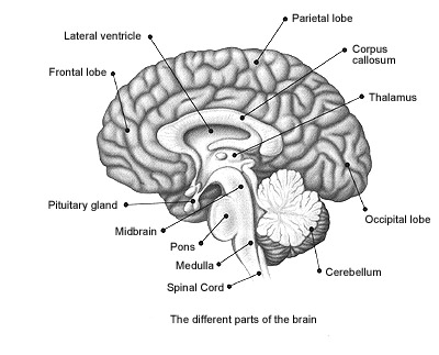
Top Level Divisions


Structure: Frontal Lobe, Parietal Lobe, Temporal Lobe, Occipital Lobe and (inside) Limbic Lobe
Description:
- Frontal Lobe - an area in the brain of humans and other mammals, located at the front of each cerebral hemisphere and positioned anterior to (in front of) the parietal lobes and above and anterior to the temporal lobes (i.e. directly behind the forehead or "temple"). It is separated from the parietal lobe by the post-central gyrus primary motor cortex, which controls voluntary movements of specific body parts associated with the precentral gyrus posteriorly, inferiorly by lateral sulcusslyvian which separates it from the temporal lobe, superiorly by the superior margin of the hemisphere and anteriorly by the frontal pole
- Temporal Lobe - a region of the cerebral cortex that is located beneath the Sylvian fissure on both cerebral hemispheres of the mammalian brain. The temporal lobe is involved in auditory perception and is home to the primary auditory cortex. It is also important for the processing of semantics in both speech and vision. The temporal lobe contains the hippocampus and plays a key role in the formation of long-term memory
- Parietal Lobe - a part of the brain positioned above (superior to) the occipital lobe and behind (posterior to) the frontal lobe. The parietal lobe integrates sensory information from different modalities, particularly determining spatial sense and navigation. For example, it comprises somatosensory cortex and the dorsal stream of the visual system. This enables regions of the parietal cortex to map objects perceived visually into body coordinate positions
- Occipital Lobe (includes Cerebellum) - the visual processing center of the mammalian brain containing most of the anatomical region of the visual cortex. The primary visual cortex is Brodmann area 17, commonly called V1 (visual one). Human V1 is located on the medial side of the occipital lobe within the calcarine sulcus; the full extent of V1 often continues onto the posterior pole of the occipital lobe. V1 is often also called striate cortex because it can be identified by a large stripe of myelin, the Stria of Gennari. Visually driven regions outside V1 are called extrastriate cortex. There are many extrastriate regions, and these are specialized for different visual tasks, such as visuospatial processing, color discrimination and motion perception. The name derives from the overlying occipital bone, which is named from the Latin oc- + caput, "back of the head"
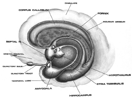
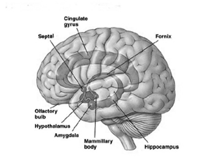
- Limbic System (includes Limbic Lobe) - a set of brain structures including the hippocampus, amygdala, anterior thalamic nuclei, septum and limbic cortex, which seemingly support a variety of functions including emotion, behavior, long term memory, and olfaction. The limbic system is the set of brain structures that forms the inner border of the cortex. In an abstract topological sense, each cortical hemisphere can be thought of as a sphere of gray matter, with a hole punched through it in the area where nerve fibers connect it to the subcortical structures of the basal forebrain. The hole is surrounded by a ring of cortical and noncortical areas that combine to make up the limbic system. The cortical components generally have fewer layers than the classical 6-layered neocortex, and are often classified as allocortex or archicortex.
Frontal Lobe

Structure: Primary Motor Cortex, Premotor Association, Frontal Eye Fields, Prefrontal Cortex, Brocas Area
- 1. Primary Motor Cortex
- function: moves individual muscules, gest - one finger, not the fist
- connected: Premotor Association
- 2. Premotor Association
- function: patterned motor movements and planning
- connected: Primary Motor Cortex , Frontal Eye Fields
- 3. Frontal Eye Fields
- function: move muscles of the eye
- connected: Premotor Association
- 4. Prefrontal Cortex:
- function: personality, judgement, complex planning, inhibition, morality
- connected: Frontal Eye Fields, Brocas Area, Insula
- 5. Brocas Area:
- function: muscles for speech
- connected: Primary Motor Cortex, Premotor Association, Frontal Eye Fields, PreFrontal Cortex, Insula
Prefrontal Cortex
Structure: Ventromedial Prefrontal Cortex, Lie Area
- 4.1 Ventromedial Prefrontal Cortex:
- lesions: people have no problem to kill
- connected: Lie
- 4.2 Lie Area:
- responsible for giving false information, lie
- connected: Ventromedial Prefrontal Cortex
- others - responsible for:
- mediating conflicting thoughts
- making choices between right and wrong or good and bad
- predicting future events
- governing social control — such as suppressing emotional or sexual urges
- the brain center most strongly implicated in qualities like sentience, human general intelligence, and personality
Temporal Lobe

Structure: Central Sulcus (valley) , Insula
- 1. Insula - in each hemisphere of the mammalian brain the insular cortex (often called insula, insulary cortex or insular lobe) is a portion of the cerebral cortex folded deep within the lateral sulcus between the temporal lobe and the frontal lobe. The cortical area overlying it towards the lateral surface of the brain is the operculum (meaning "lid"). The opercula are formed from parts of the enclosing frontal, temporal and parietal lobes. The insular cortex is divided into two parts: the larger anterior insula and the smaller posterior insula in which more than a dozen field areas have been identified. The insulae play a role in diverse functions usually linked to emotion or the regulation of the body's homeostasis. These functions include perception, motor control, self-awareness, cognitive functioning, and interpersonal experience. Related to these it is involved in psychopathology.
- function: habits, visceral sensations, deep in the valley
- lesions: alleviate habits
- connected: Prefrontal Cortex, Brocas Area, Primary Auditory Cortex
- 2. Primary Auditory Cortex
- function: basic hearing - pitch, rhythm, loudness
- connected: Insula
- 3. Auditory Association Cortex
- function: actual perception of sound - how do you respond emotionally
- connected: Primary Auditory Cortex
Parietal Lobe
Structure: Primary Somatosensory Cortex, Somatosensory Associaion, Gestatory
- 1. Primary Somatosensory Cortex
- function: basic sensations of the body
- connected: Primary Motor Cortex, Gestatory, Somatosensory Associaion
- 2. Somatosensory Associaion
- function: size, texture and relationship
- connected: Primary Somatosensory Cortex, Gestatory,
- 3. Gestatory
- function: taste
- connected: Primary Auditory, Primary Motor Cortex, Primary Somatosensory Cortex, Somatosensory Association
- 4. Werniche's Area
- function: sound and words
- 5. General Interpretation Area:
- function: opinions, can I fit this in here
Occipital Lobe
Structure: Primary Visual Cortex
- Primary Visual Cortex
- function: simple lines and shapes
- connected: Visual Associations
- Visual Associations
- function: color, form and movement
- movement part area: MT - lesions lead to see life in strobe, no motion
- connected: Primary Visual Cortex
- Fusiform Gyris:
- lesions: you cas see but you cannot recognise faces
- connected: Visual Associations
- Cerebellum
- function: complex motion behavior, sequential thinking
- lesions: you cannot drive bike
- connected: Primary Visual Cortex, Visual Associations, Fusiform Gyris
Limbic Lobe


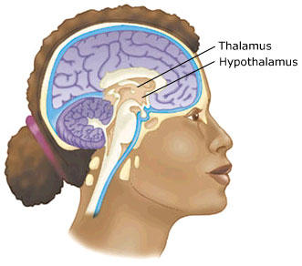
Structure: Thalamus, Choroid Plexus, Pineal Gland, Corpus Callosum, Hypothalamus, Pituitary
- 1. Thalamus
- function: sorts out the information, groups and prioritises information
- connected: Insula
- lesions: cannot pick out important things, cannot keep eye contact, overwhelmed by sensations
- 2. Choroid Plexus
- function: makes CSF
- details: Cerebrospinal fluid (CSF), Liquor cerebrospinalis, is a clear bodily fluid that occupies the subarachnoid space and the ventricular system around and inside the brain and spinal cord. In essence, the brain "floats" in it. CSF proves a basic mechanical and immunological protection to the brain inside the skull.
- 3. Pineal Gland
- function: wake-sleep cycles
- details: It produces the serotonin derivative melatonin, a hormone that affects the modulation of wake/sleep patterns and seasonal functions. Its shape resembles a tiny pine cone (hence its name), and it is located near the center of the brain, between the two hemispheres, tucked in a groove where the two rounded thalamic bodies join.
- 4. Corpus Callosum
- function: connects left and right hemispheres
- lesions: split brain - 1) if shown to right eye, can identify but not describe function (right eye go to left brain, left brain is responsible for language), 2) if shown to left eye, can describe but cannot name
- 5. Hypothalamus
- function: blood pressure, hunger, breathing, body temp, hormon cycles
- 6. Pituitary (pituitary gland and pituitary fossa, hypophysis)
- function: synthesizes and secretes the important endocrine hormones - Human Growth, Adrenalin, for sexual development and reproductive function